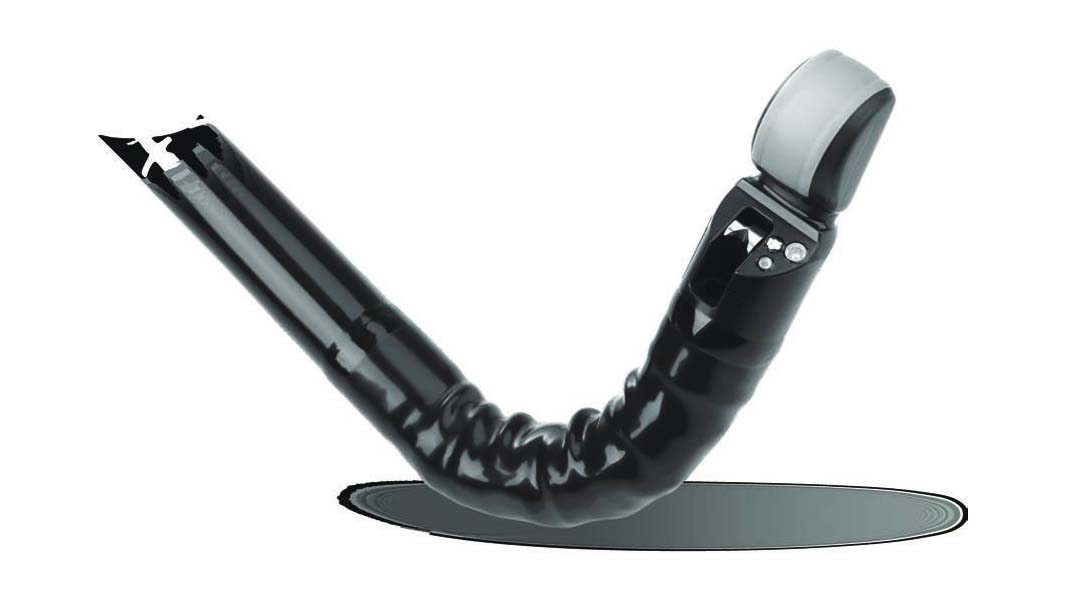
4.0-mm Instrument channel
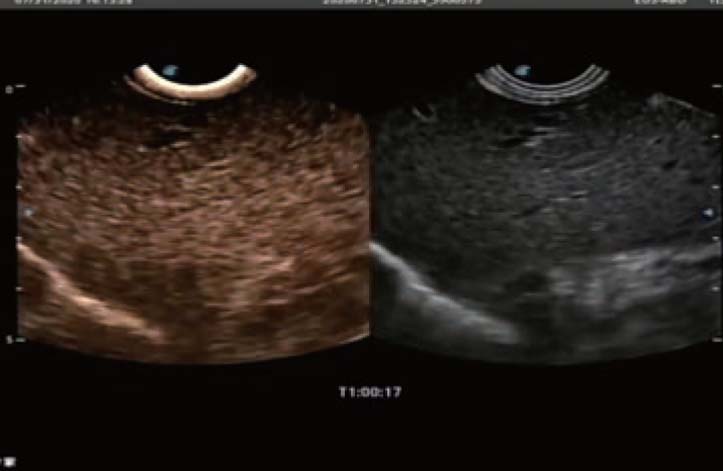
Linear Array Echoendoscope
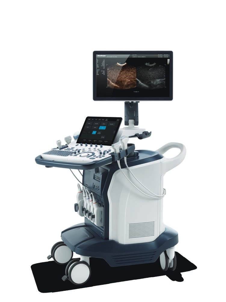
EG-UC5T
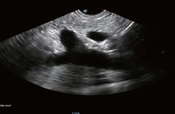
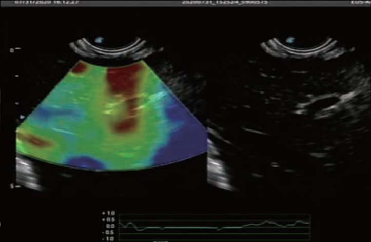
Pushing the boundaries of Therapeutic excellenceReaping the benefits of decades of ultrasound technology know-how, combined with our endorsed endoscope design, EG-UCS5T widens staging and therapeutic options, fulfils and exceeds the most sought-after requirement from your medical team, levelling up the vision and achievement behind the wall of the Gl tract.